What Is MRI?
Magnetic Resonance Imaging, commonly referred to as MRI, is a primary imaging modality used in radiology. MRI uses a magnetic field and radio frequencies to develop detailed, high-resolution images of organs and tissues. MRI creates cross sectional images of the study, allowing physicians to view cross sections of the area of interest to find the problem at hand.How does an MRI machine work?
On an atomic level, an MRI machine introduces a magnetic field to the protons which are found in the human body. The protons align with this magnetic field, which is then manipulated by adding radio frequencies that realign the protons in a different direction than the magnetic field. One these frequencies stop, the protons realign and release electromagnetic energy. This stimulates the area of interest in the body, which allows the machine to take images of the portion.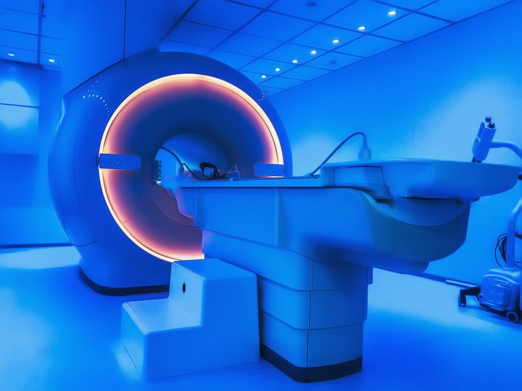
MRI Over Other Modalities
Magnetic Resonance imaging is a non-invasive imaging procedure that allows physicians to analyze organs, tissues, and bones in a more in depth manner than other imaging modalities.Advantages of MRI over other Primary Modalities:
- MRI does not use ionizing radiation unlike CT, X-rays, and PET scans. MRI is often seen as the safer and generally more superior alternative to these modalities due to its use of magnetic and computer radiography to take computerized images instead of relying on radiation.
- MRI is able to create high definition cross sectional views of the area of interest that other modalities cannot do, physicians prefer MRI images for this reason
- MRI is able to view soft tissues such as muscles, ligaments, and tendons in addition to bones, which is very limited or non-existent for the other modalities.
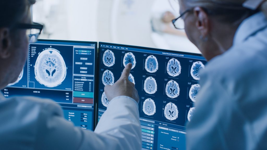
- Injuries involving the brain and spinal cord are usually assessed through MRI because of the clear, high quality images, which makes it superior to CT scans
- MRI is able to differentiate between fat, water, muscle, and soft tissue in a much more capable manner than CT
MRI History
Initially, the concept of Magnetic Resonance Radiography was established out of research of nuclear physics during the Second World War. Scientists such as Isidor Rabi, Enrico Fermi, and Ernest Lawrence developed the concept of Nuclear Magnetic Resonance, a phenomenon in which a powerful magnetic field comes into contact with a weak oscillating magnetic field, creating an electromagnetic field that allow nuclei of atoms to become aligned with the magnetic field.
Later on, this concept was adopted by researchers such as Raymond Damadian and Paul Lauterbur, in which they applied this phenomena in order to create imaging technology. Using the NMR phenomena and radio frequency technology, researchers were able to create a new imaging device that was able to do far more than the existing X-ray technology. In 1977, Dr. Damadian conducted the first MRI exam, and it was hailed a huge success.
By the 1980s, MRI became a standard modality in the field of Radiology and was a common sight to see in many Hospitals, imaging centers, and research institutions. With the introduction of computer systems and computerized imaging, MRI machines became even easier to operate and were ideal for radiologists and other physicians due to the technological advantages that the MRI provided them.
Today, MRI is an industry standard imaging modality that has an important role in the medical field due to its versatility and technological capabilities
